Description
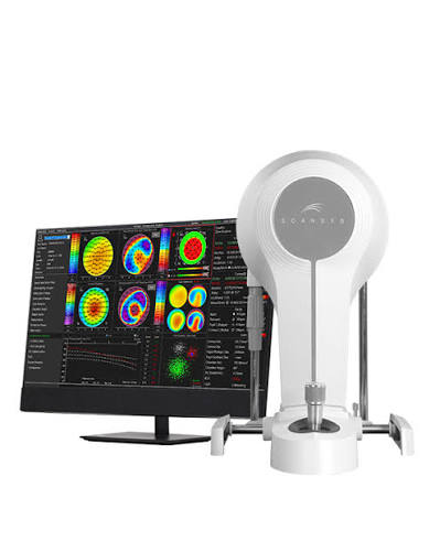
The Scansys TA517 Pro Anterior Segment Analyzer is a state-of-the-art diagnostic instrument designed for in-depth analysis of the ocular anterior segment. Utilizing ultra-high-definition Scheimpflug imaging technology, it delivers precise, high-resolution images and data essential for comprehensive ocular evaluation. The device features a robust array of diagnostic capabilities, including the capture of up to 662,400 data analysis points, enabling exceptional measurement accuracy and detail.
Engineered with advanced optics and a refined algorithmic framework, the TA517 Pro supports a wide spectrum of clinical applications from routine diagnostics to complex surgical planning. Its sophisticated software allows for the simulation and analysis of various optical phenomena, such as wavefront aberrations and corneal topography, providing critical insights for enhanced patient outcomes. The system’s extensive data integration capabilities, combined with its user-friendly interface, streamline the diagnostic process and facilitate precise patient management across a range of ophthalmic and optometric scenarios.
Keratoconus Diagnosis
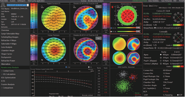
With its advanced keratoconus analysis capabilities, the TA517 Pro provides detailed corneal thickness spatial profiles and refractive maps. It incorporates indices like the Keratoconus Index (KCI) and Keratoconus Probability (KCP) to deliver precise evaluations. This feature is essential for early detection, monitoring disease progression, and guiding treatment plans, thus improving patient management outcomes.
Refractive Cataract Surgery
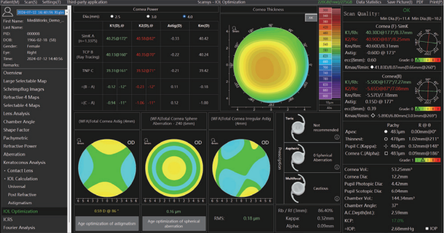
The Scansys TA517 Pro is indispensable in refractive cataract surgery, offering precise IOL power calculations with new formulas such as Pearl-DGS and Pearl-DGS Pro. These algorithms account for post-refractive surgery scenarios, enhancing the accuracy of IOL power predictions. The system’s ability to simulate surgical outcomes and optimize IOL selection ensures superior visual results and patient satisfaction.
Implantable Collamer Lens (ICL) Surgery
Supporting ICL surgery, the analyzer offers comprehensive preoperative and postoperative assessments. It analyzes corneal diameter, curvature, thickness, and anterior chamber depth to recommend the optimal ICL size and predict postoperative vault. The device’s high-definition imaging capabilities ensure precise evaluation, facilitating safer and more effective surgical outcomes.
Intrastromal Corneal Ring Segment (ICRS) Surgery
The TA517 Pro provides critical data for ICRS implantation, focusing on improving predictability and reducing complications. By analyzing the corneal stromal ring tunnels, both through manual and femtosecond laser dissection, it aids in crafting precise surgical plans. This level of detail is crucial for achieving optimal patient outcomes and enhancing surgical success rates.
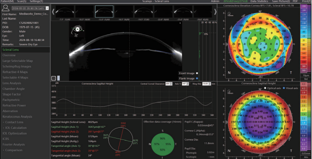
Featuring an innovative contact lens fitting module, the TA517 Pro supports scleral, RGP, and soft lens fittings with unmatched accuracy. It automatically calculates fitting parameters using corneal topography data, and simulates fluorescein patterns under different conditions, optimizing the fitting process. This reduces trial and error, enhances patient comfort, and significantly improves fitting efficiency, particularly for complex cases like orthokeratology.
Visual Quality Analysis
The Scansys TA517 Pro excels in visual quality analysis by leveraging advanced algorithms to generate detailed wavefront aberrations up to the 7th order. This allows for precise simulation of visual outcomes, both pre- and post-surgery. The system visualizes Point Spread Function (PSF) and Modulation Transfer Function (MTF), providing comprehensive insights into the patient’s visual quality, which is crucial for tailored vision correction strategies.
Glaucoma Screening
The TA517 Pro is equipped with sophisticated tools for glaucoma screening, including anterior chamber angle analysis and intraocular pressure (IOP) correction formulas based on corneal thickness. It measures parameters such as Angle Opening Distance (AOD) and Trabecular-Iris Space Area (TISA), providing a comprehensive assessment of the anterior chamber. This facilitates early detection and effective management of glaucoma, aiding in the prevention of vision loss.